
Internal Anatomy Of The Eye Labeled Life Educations
Apr. 29, 2023 To understand the diseases and conditions that can affect the eye, it helps to understand basic eye anatomy. Here is a tour of the eye starting from the outside, going in through the front and working to the back. Eye Anatomy: Parts of the Eye Outside the Eyeball The eye sits in a protective bony socket called the orbit.
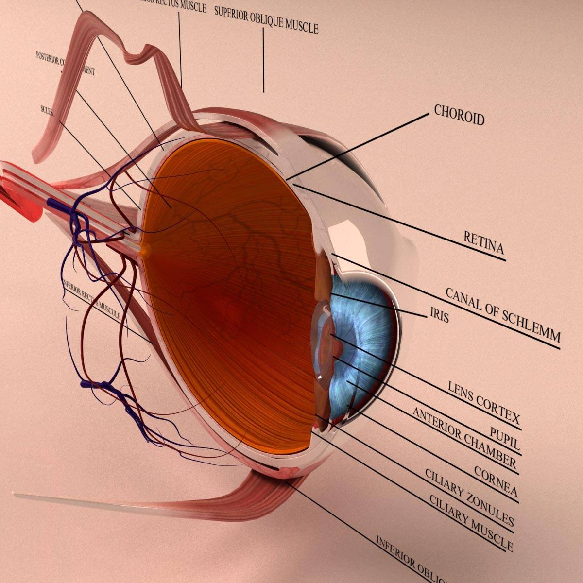
Anatomy Human Eye Cross Section 3D Model Kezan's Portfolio
Definition. an oval yellowish area of the retina surrounding the fovea near the center of the retina in the eye, which is the region of keenest vision. Location. Term. fovea centralis. Definition. a small depression in the macula with an abundance of cones; it is the area of clearest vision. Location.

Image result for Eye Model Labeled Apuntes de clase, Maquetas, Escuela
Eyes Anterior chamber. The front section of the eye's interior where aqueous humor flows in and out, providing nourishment to the eye. Aqueous humor. The clear watery fluid in the front of the eyeball. Blood vessels. Tubes (arteries and veins) that carry blood to and from the eye. Caruncle.

Human Eye Discovering DNA
Anatomy of the Extraocular Muscles Interactive ophthalmic figures for medical student education illustrate concepts in eye anatomy and functions in an engaging format.

Inner Eye Structure Anatomy Anatomy Body Gallery Eye anatomy
Our extensive selection of eye models includes general human eye models, detail specific medical eye models, and conditions of the eye anatomy models . Many of our human eye models are attached to a base allowing for easy rotation and viewing. Enlarged sizes are great for beginning optometry students or even undergraduate and secondary students.

Eye Model Labeled Bing Images Eye anatomy, Eye health, Eye sight
Human eye models are widely used in teaching of Human Anatomy in schools and colleges. Eye models and charts are also used to teach optometrists and opticians and also for patient education. All eye models are of medical quality. Pathological Human Eye Model - 3B Smart Anatomy $ 405.00 Item: 1017230 [F17]

Human Eye Diagrams with the Unlabeled 101 Diagrams
Eye Models. Click on Label for the labeled model. Back to Nervous System. Eye with Extrinsic Muscles. Large Eye (lateral) Large Eye with Choroid Coat. Label. Label. Label.

Eye Model Labeled Bing Images Biology Pinterest Eye, Anatomy
A mystical journey through the major landmarks of the eye.Diagram: http://droualb.faculty.mjc.edu/Lecture%20Notes/Unit%203/Extrinsic_eye_muscles_3.jpg

February 2014 optometry blog
Support and Taste cells. This page titled 13.6: MODELS- Eye, Ear, Torso, Mid-Sagittal Head and Cochlea is shared under a CC BY-NC-SA 4.0 license and was authored, remixed, and/or curated by Laird C. Sheldahl via source content that was edited to the style and standards of the LibreTexts platform; a detailed edit history is available upon request.

eye diagram Discovery Eye Foundation
Realistic cross-section of the Eye. This 3D model can be licensed from MotionCow by Educators, 3D Artists and App Developers.
SCB209 Lab3 Natural Sciences Open Educational Resources
Labelling the eye. Use this interactive to label different parts of the human eye. Drag and drop the text labels onto the boxes next to the diagram. Selecting or hovering over a box will highlight each area in the diagram. The human eye has several structures that enable entering light energy to be converted to electrochemical energy.

Eye Model Labeled Bing Images Anatomy Pinterest Med school
choroid. ciliary nerves. retina. ciliary part of retina. macula and fovea centralis. optic disc. lens. vitreous body. Study with Quizlet and memorize flashcards containing terms like superior rectus, inferior rectus, medial rectus and more.

Eye Model Labeled Bing Images Biology Pinterest Nursing programs
PP2022AMB4385 Learn basic eye anatomy with interactive 3D models, view video tutorials of common refractive errors, and see how contact lenses can help give you clear vision.

3d model human eye section Eye anatomy, Medical anatomy, Eyeball anatomy
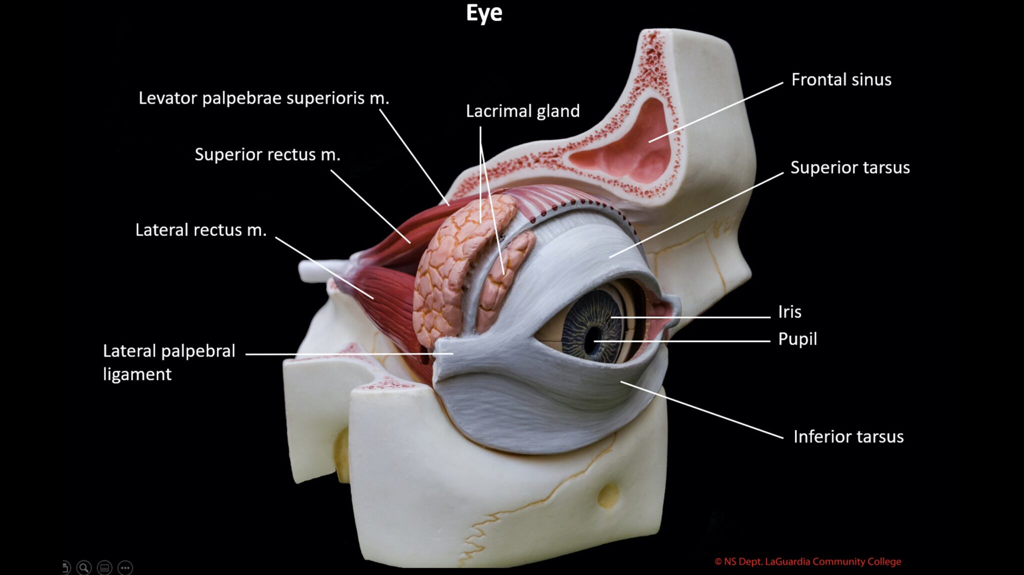
External Right Eye Model 1. Frontal Bone 9. Superior Rectus 2. Nasal Bone 10. Trochlea of Superior Oblique 3. Maxillary Bone 11. Lacrimal Gland 4. Lacrimal Bone 12. Sclera 5. Zygomatic Bone 13. Iris 6. Inferior Rectus 14. Pupil 7. Inferior Oblique 15. Nasolacrimal Duct 8. Lateral Rectus 16.…

anatomy eye model labeled Tempat untuk Dikunjungi Pinterest Eye
Human Eye Anatomy Model. 3B Scientific. Retail Price $382.00. Today's Price $314.00. Learn about the human eye anatomy in-depth with our collection of eye anatomical models and charts. Whether you are an optometrist or professor of human anatomy, use our detailed standard eye charts for sale or 3D model of the eye for classroom teaching or.

Anatomical Eye Model ANATOMY
Hello, in this video I will explain in detail the anatomical landmarks of the human eye. Thanks for watching, don't forget to like and subscribe and leave a.